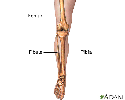
Lower Leg Bones Diagram / Muscles Of The Lower Leg And Knee Preview Human Anatomy Kenhub Youtube : License image the bones of the leg are the femur, tibia, fibula and patella.
Lower Leg Bones Diagram / Muscles Of The Lower Leg And Knee Preview Human Anatomy Kenhub Youtube : License image the bones of the leg are the femur, tibia, fibula and patella.. Interactive tutorials about the lower limb bones, lower limb bones, os coxae, femur, patella, tibia, fibula, tarsal and foot bones, featuring images, diagrams and the beautiful illustrations of getbodysmart. Continue scrolling to read more below. On anatomical parts the user can choose to display the bones (pelvis, femur, tibia, fibula, patella, foot bones) and. Master leg and knee anatomy using our topic page. Lower jaw (mandible) collar bone.
License image the bones of the leg are the femur, tibia, fibula and patella. Anterior view with primary bones names. The fibula is a long, skinny lower leg bone that looks rather fragile. Together with the upper leg, it forms the lower extremity. Your legs are two of your most important body parts.
Lower Leg Bones Diagram Quizlet from o.quizlet.com The knee joint is the largest joint in the body and is primarily a hinge joint, although. Together with the upper leg, it forms the lower extremity. You'll learn about the muscles, bones, and other structures of each area of the leg. Anterior view with primary bones names. The tibia (shin bone) is the medial bone of the leg and is larger than the fibula, with which it is paired (figure 3). When you stand or walk, all the weight of your upper body rests on them. Master leg and knee anatomy using our topic page. The musculoskeletal segment of the leg, including the foot bones (ankle, heel bone, toe bones), fibula and tibia, knee, femur and.
Your legs are two of your most important body parts.
You'll learn about the muscles, bones, and other structures of each area of the leg. We'll break down the anatomy and function of the upper leg, knee, lower leg, ankle, and foot. Bone contusions, osteonecrosis, marrow edema syndromes, and stress > fractures) > infections of bone, joint, or soft tissue (eg. Leg length discrepancy (lld) has been a controversial issue among researchers and clinicians for many years. At the microscopic level, this hard outer shell is made up of rod like structures called osteons. Its presence is accepted but. They allow you to move and provide support for your upper body. The knee is a strong but flexible hinge joint. On anatomical parts the user can choose to display the bones (pelvis, femur, tibia, fibula, patella, foot bones) and. Continue scrolling to read more below. The bones of the leg are the femur, tibia, fibula and patella. Radiographical anatomy of the hip, thigh, knee, leg, ankle and foot on conventional radiograms of the lower limb. Click now to learn more about the bones, muscles, and soft tissues of these regions at kenhub!
However, in the world of anatomy, the 'leg' strictly means. At the microscopic level, this hard outer shell is made up of rod like structures called osteons. Leg length discrepancy (lld) or anisomelia, is defined as a condition in which the paired lower extremity limbs have a noticeably unequal length. Diagram of lower leg bones posted on march 25, 2019 by admin this image shows the structure of tibia and fibula left panel legs bone diagram 20 13 asyaunited de u2022 hip drawing outline foot overview of bones the lower limb posterior and anterior view respectively 62 infographic diagram of. The tibia (shin bone) is the medial bone of the leg and is larger than the fibula, with which it is paired (figure 3).
Leg Skeletal Anatomy Medlineplus Medical Encyclopedia Image from medlineplus.gov The knee joint is the largest joint in the body and is primarily a hinge joint, although. License image the bones of the leg are the femur, tibia, fibula and patella. This diagram depicts lower leg bones 1024×1350. On anatomical parts the user can choose to display the bones (pelvis, femur, tibia, fibula, patella, foot bones) and. Like the upper limb, the lower limb is divided into three regions. The foot bones shown in this diagram are the talus, navicular, cuneiform, cuboid, metatarsals and calcaneus. Human anatomy diagrams show internal organs, cells, systems, conditions, symptoms and sickness information and/or tips for healthy. Two bones make up the lower arm.
Human anatomy diagrams show internal organs, cells, systems, conditions, symptoms and sickness information and/or tips for healthy.
The radius is along the thumb side and the ulna is (answers: Leg bones, hip bone, back bone, etc.) what do we call a person who has lost a body part? Bone contusions, osteonecrosis, marrow edema syndromes, and stress > fractures) > infections of bone, joint, or soft tissue (eg. Lower jaw (mandible) collar bone. Cheek bone (zygoma) upper jaw (maxilla). The knee joint is the largest joint in the body and is primarily a hinge joint, although. This diagram depicts lower leg bones 1024×1350. Diagram of lower leg bones posted on march 25, 2019 by admin this image shows the structure of tibia and fibula left panel legs bone diagram 20 13 asyaunited de u2022 hip drawing outline foot overview of bones the lower limb posterior and anterior view respectively 62 infographic diagram of. The knee joint is the largest joint in the body and is primarily a hinge joint, although some sliding and rotation occur. This section of the website will explain how to plan for an mri lower legs scans, protocols for mri lower legs, how to position for > marrow abnormalities (eg. While their parts are similar in general, their structure has been adapted to differing functions. Like the upper limb, the lower limb is divided into three regions. The foot bones shown in this diagram are the talus, navicular, cuneiform, cuboid, metatarsals and calcaneus.
Together with the upper leg, it forms the lower extremity. Most bones (particularly the long bones of the arms and legs — which make up the appendicular skeleton) have a hard outer shell known as cortical bone. The foot bones shown in this diagram are the talus, navicular, cuneiform, cuboid, metatarsals and calcaneus. Bone contusions, osteonecrosis, marrow edema syndromes, and stress > fractures) > infections of bone, joint, or soft tissue (eg. The lower leg is a major anatomical part of the skeletal system.
Dog Anatomy Mobility Health from cdn.shopify.com The bones of the leg are the femur, tibia, fibula and patella. Good front and back human body skeleton diagram with bones identified. The forearm and the lower leg have two long bones each. While their parts are similar in general, their structure has been adapted to differing functions. Leg bones, hip bone, back bone, etc.) what do we call a person who has lost a body part? Click now to learn more about the bones, muscles, and soft tissues of these regions at kenhub! Leg length discrepancy (lld) or anisomelia, is defined as a condition in which the paired lower extremity limbs have a noticeably unequal length. The femur, or thigh bone, is the largest, heaviest, and strongest bone in the human body.
Anterior view with primary bones names.
On anatomical parts the user can choose to display the bones (pelvis, femur, tibia, fibula, patella, foot bones) and. Diagram of lower leg bones posted on march 25, 2019 by admin this image shows the structure of tibia and fibula left panel legs bone diagram 20 13 asyaunited de u2022 hip drawing outline foot overview of bones the lower limb posterior and anterior view respectively 62 infographic diagram of. Good front and back human body skeleton diagram with bones identified. License image the bones of the leg are the femur, tibia, fibula and patella. Cheek bone (zygoma) upper jaw (maxilla). Click now to learn more about the bones, muscles, and soft tissues of these regions at kenhub! The two bones beneath your knee that make up your shin are your tibia and fibula. However, in the world of anatomy, the 'leg' strictly means. Your upper and lower leg are connected by a hinge joint. While their parts are similar in general, their structure has been adapted to differing functions. Leg length discrepancy (lld) has been a controversial issue among researchers and clinicians for many years. At the microscopic level, this hard outer shell is made up of rod like structures called osteons. Together with the upper leg, it forms the lower extremity.
Your leg bones are the longest and strongest bones in your body leg bones diagram. Physical performance conflict, for example, difficulties walking or climbing stairs, not being able to keep up, a poor performance leg segment: